1 / 5
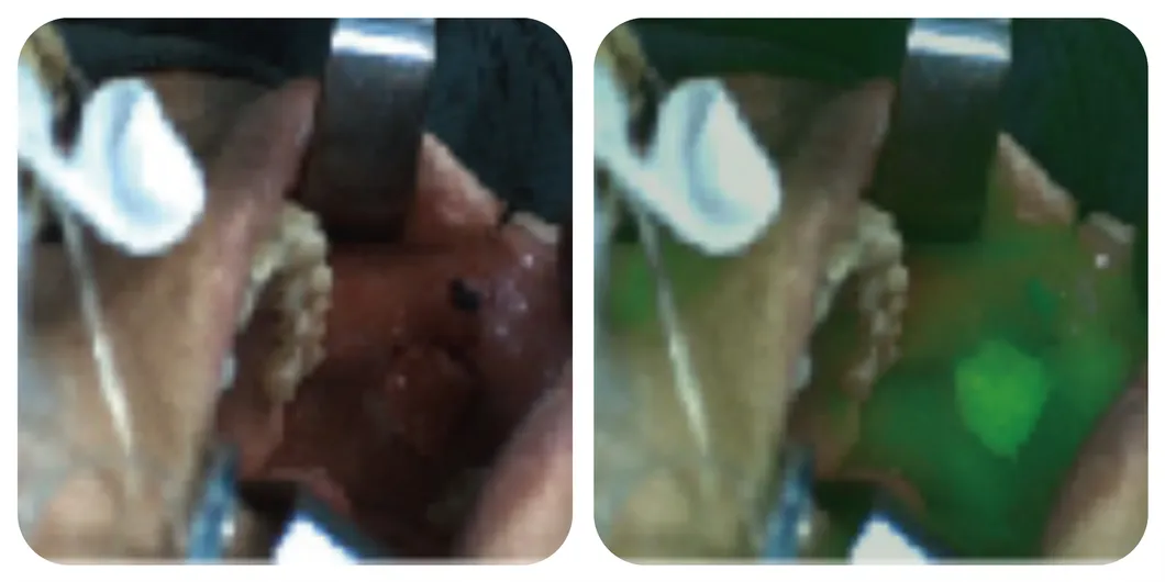
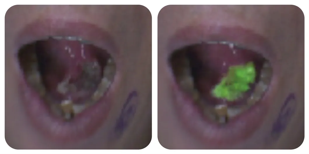
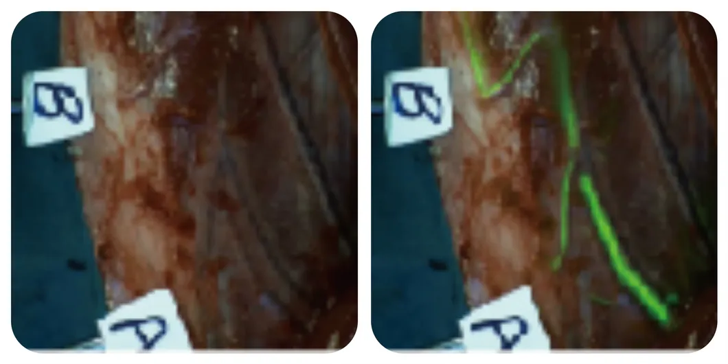
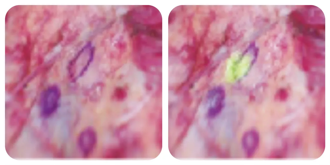
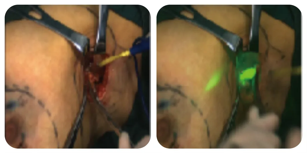
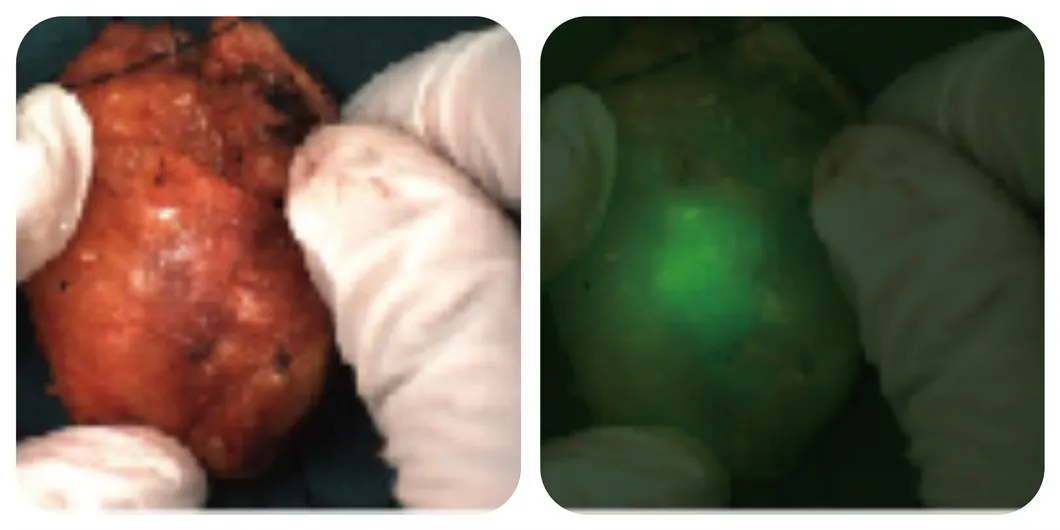
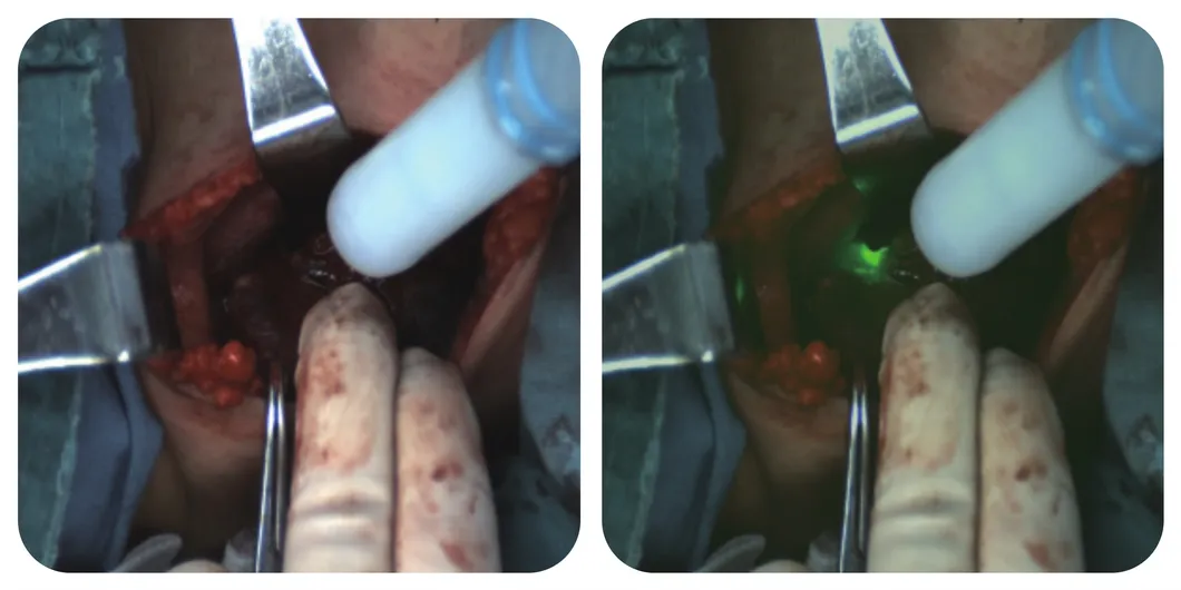
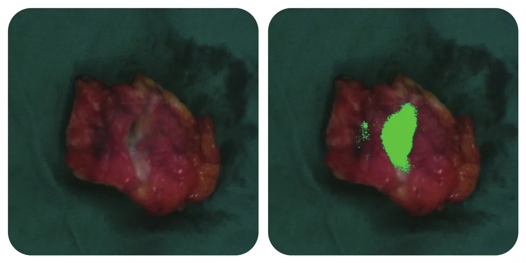
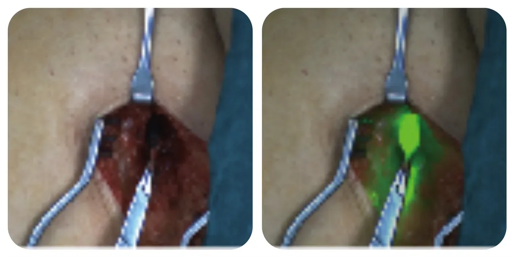
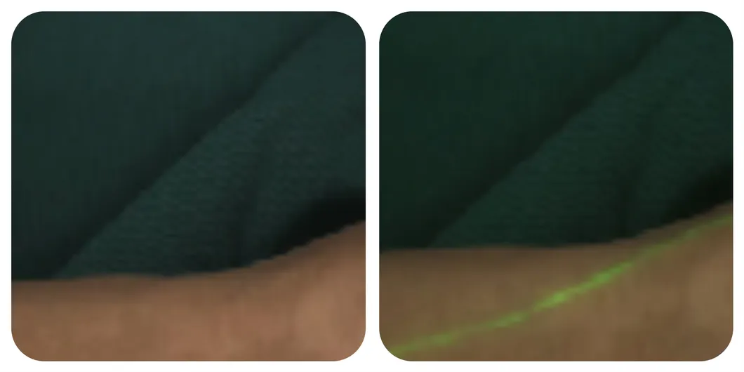
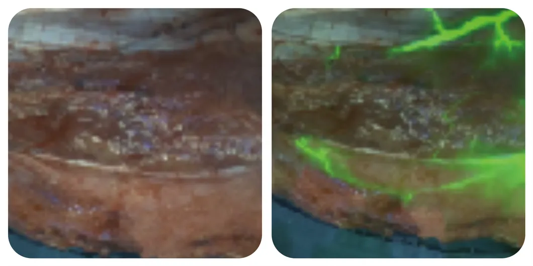
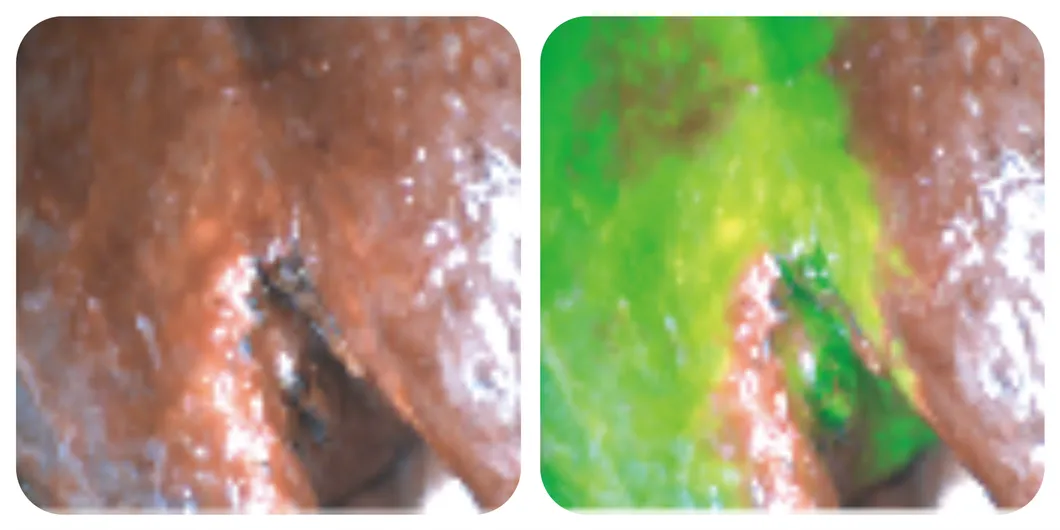
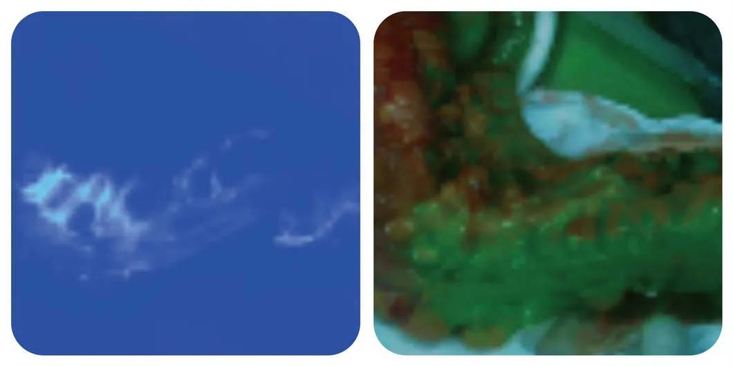
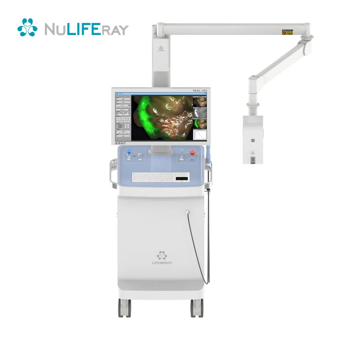
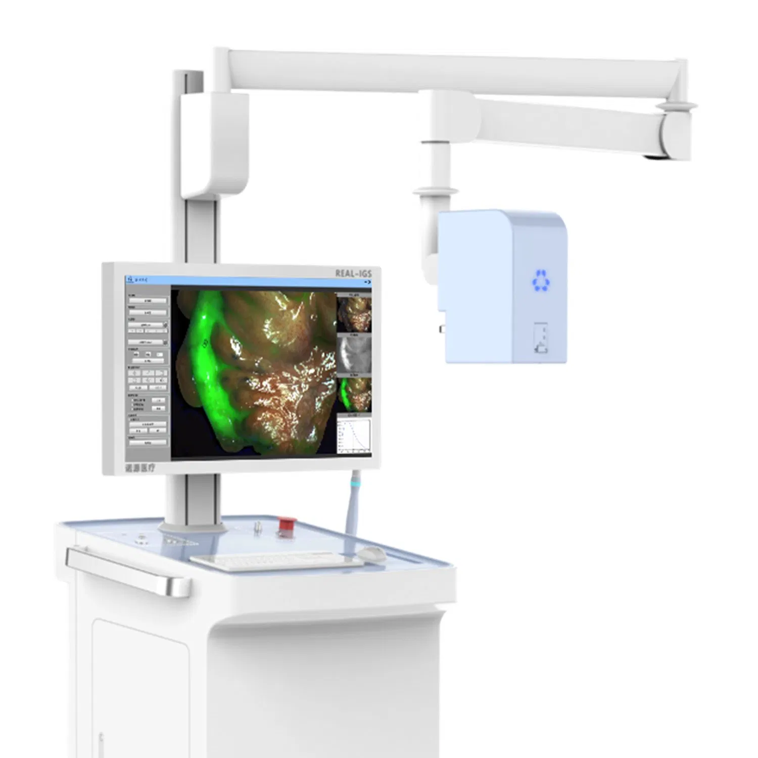
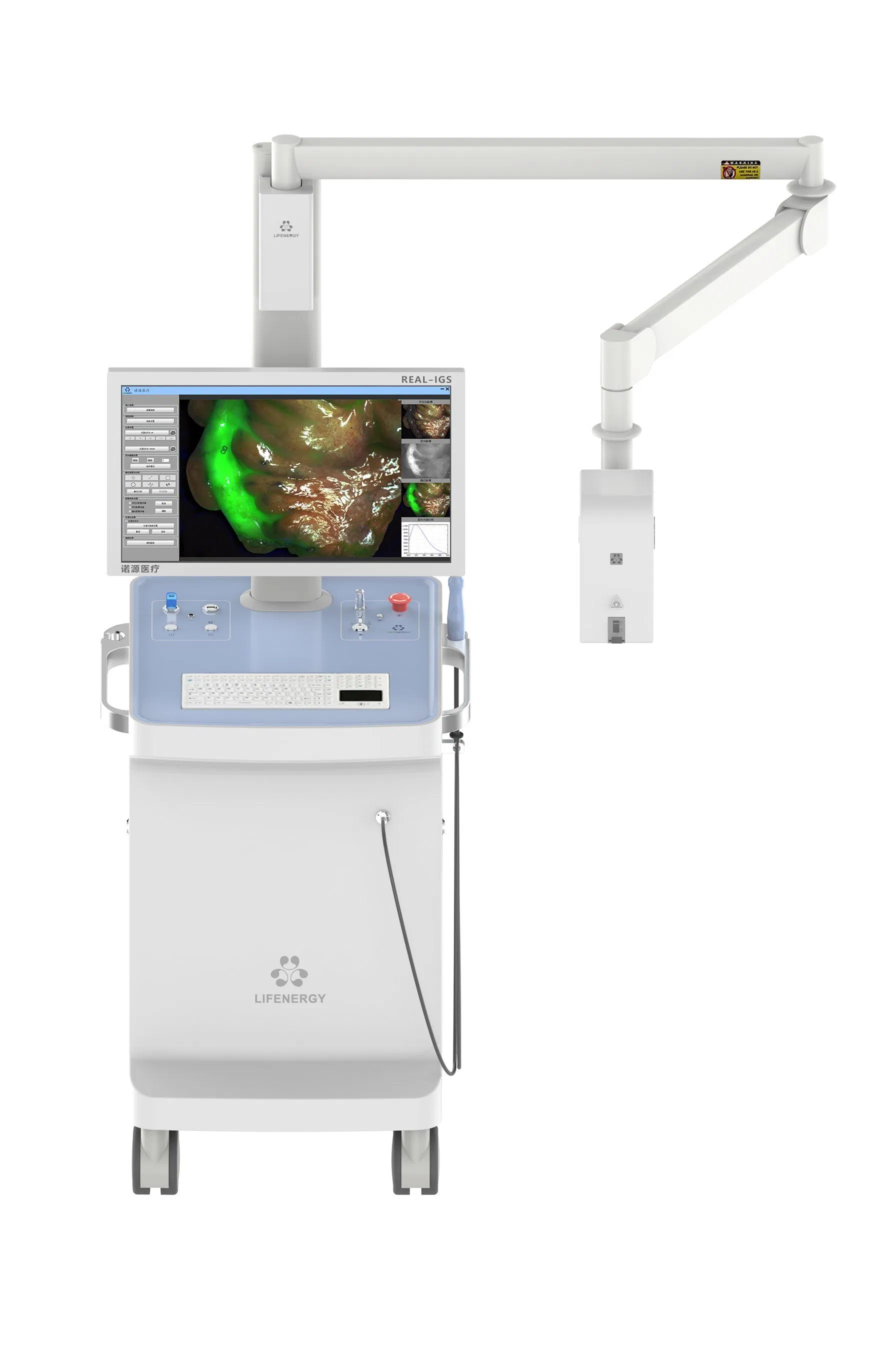
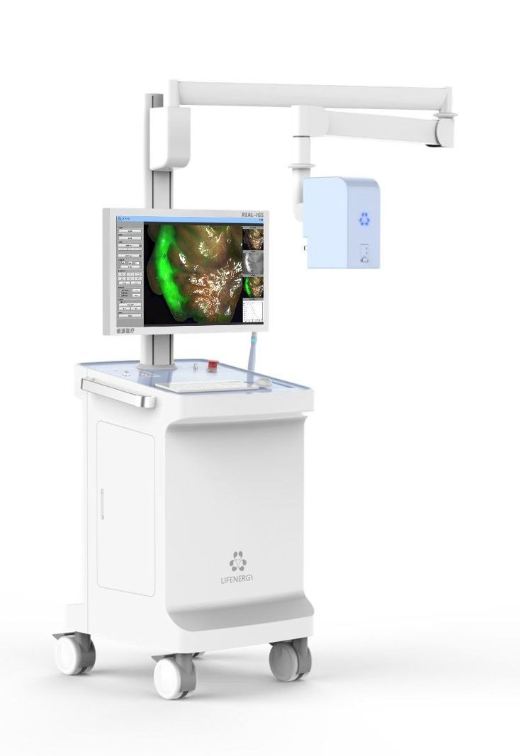
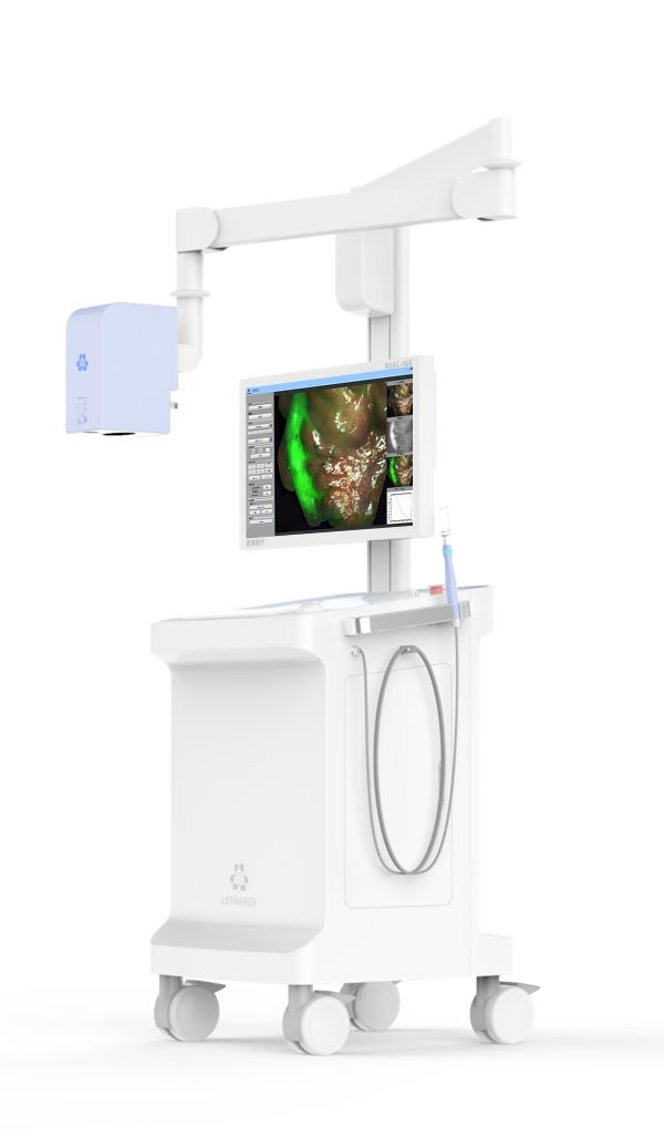
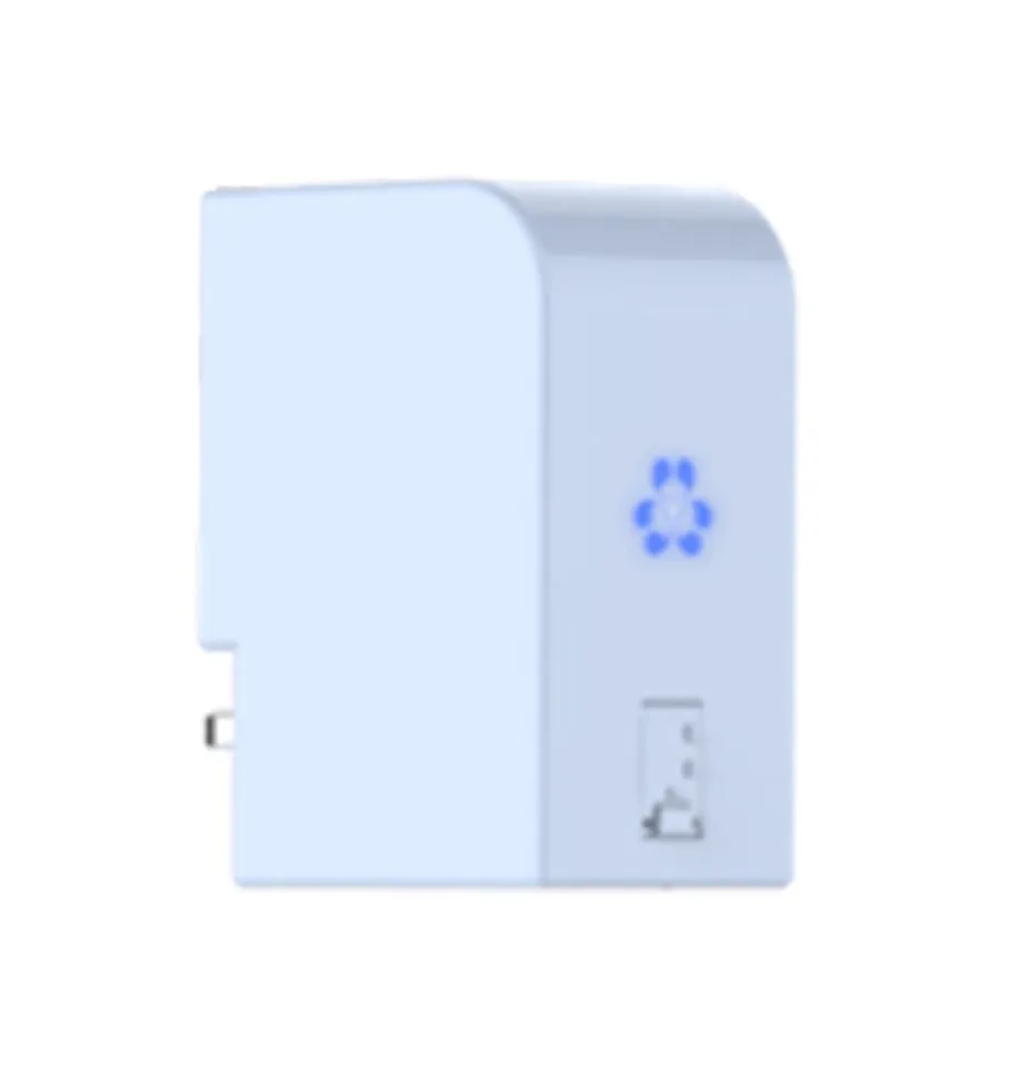
The FLI-10B surgical fluorescence imaging system is a surgical guidance system that uses a drug-device combination approach. It employs indocyanine green (ICG) as a fluorescence probe, relying on "ultra-high sensitivity" and combining the optical properties of ICG in submillimeter-sized tumors to provide surgeons with high-definition visible light, fluorescence imaging, and quantitative data for diagnostic information during tumor surgery.
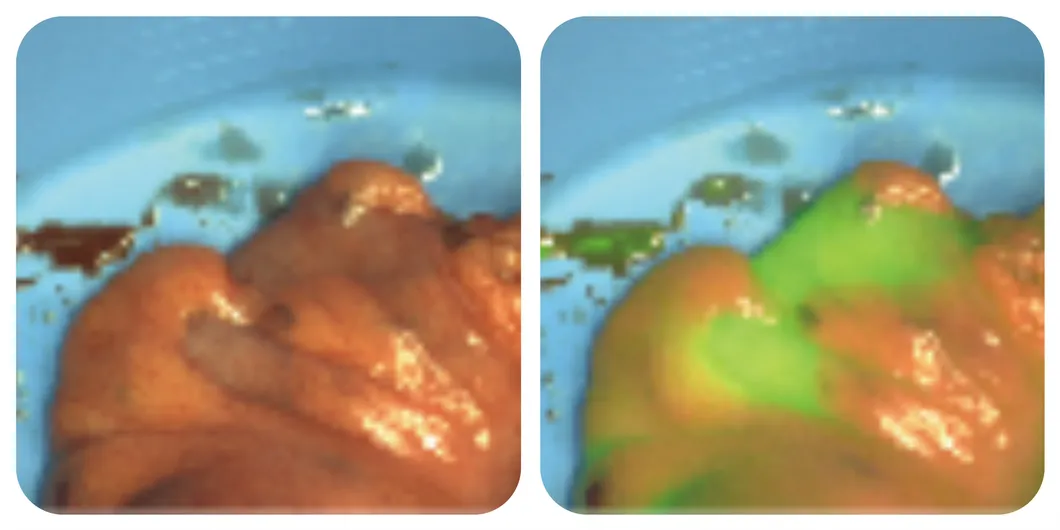
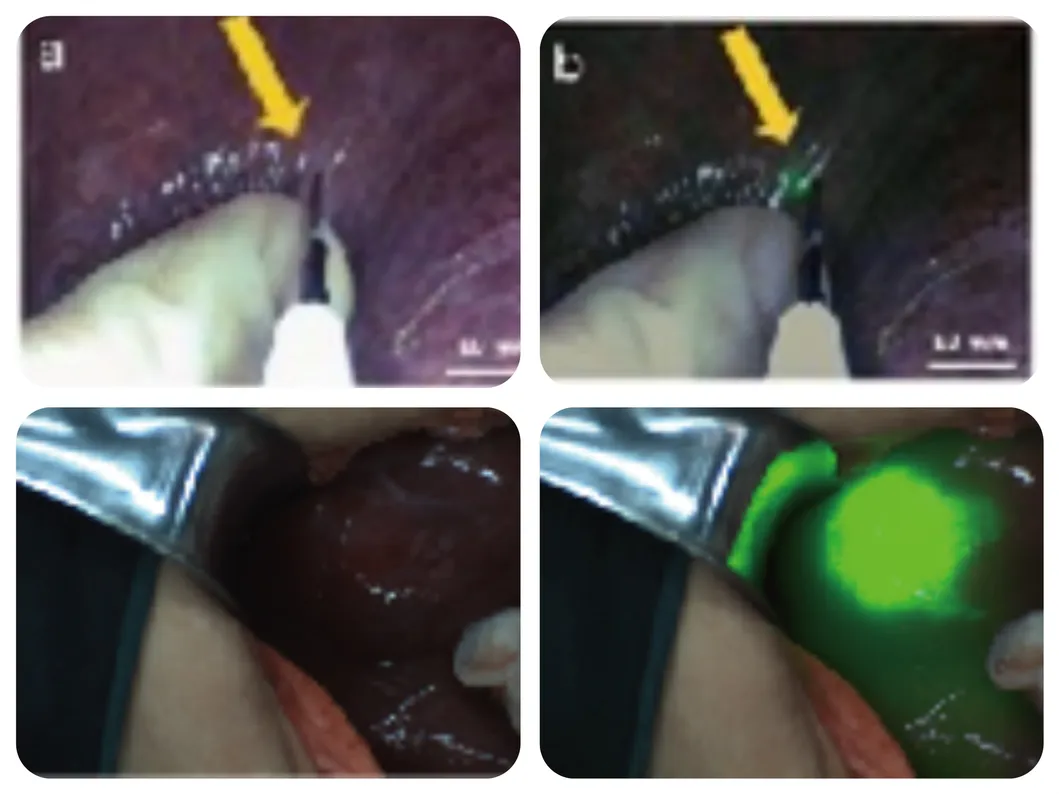
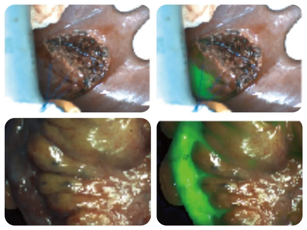
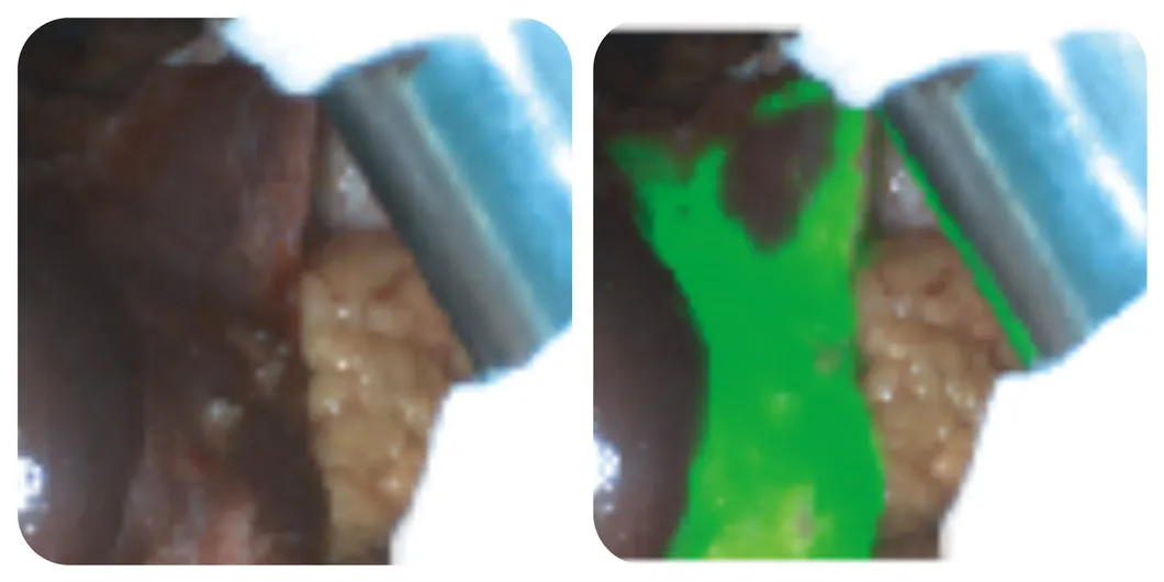
It is suitable for real-time observation of tissues (such as tumor tissue, margin tissue), blood supply (free skin flap), lymph nodes (sentinel lymph nodes, regional lymph nodes), anatomical structures (liver segments, gallbladder, lung segments), to make more accurate medical judgments, optimize surgical plans, and evaluate treatment efficacy.
| Project | Content |
|---|---|
| Number of Camera Chips | 2CMOS |
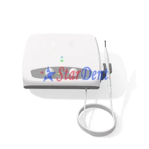
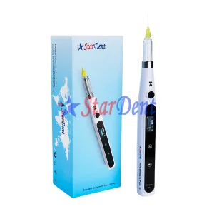
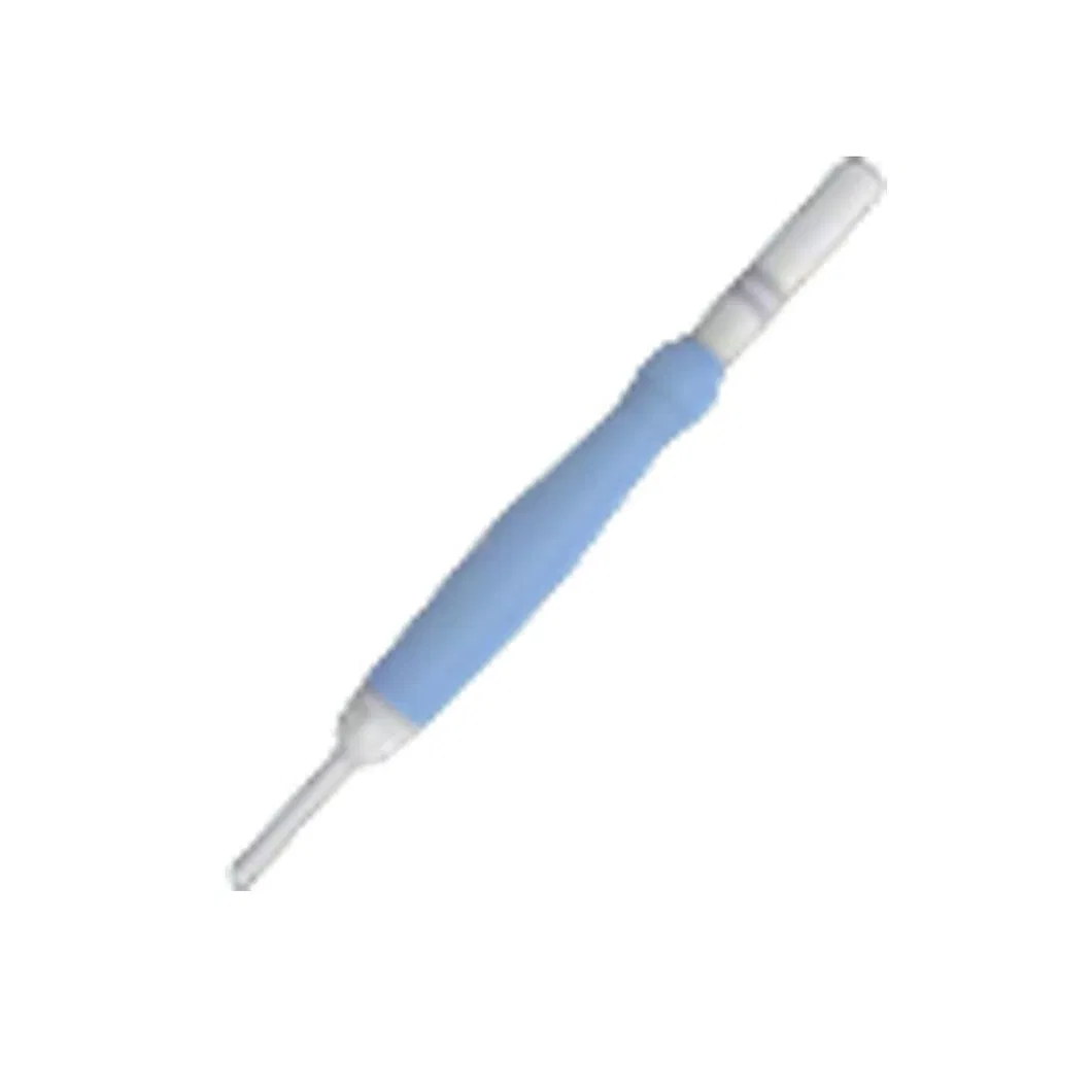
| Convenient Handheld Probe | Handheld Spectral Quantitative Analysis Probe |
| Special Fluorescence Development | AI Assisted Boundary Sharpening |
| Image Mode | 7 Types: White Light/Fluorescence/Fusion/Multimode/ColorGrading/Quantification/Spectroscopy |
| Laser Wavelength | 785nm |
| Is There a Workstation Available | Yes |
| Workstation Function | Fluorescence Intensity & Spectral Quantitative Analysis |
| Fluorescence Detection Limit | 10-12M/L |
| Lens Zoom Factor | 4 Times |
| Camera Working Distance | Recommended 10-25cm |
| Focus Mode | Electric Focus |
| White Balance Method | Manual White Balance |
| Laser Grade | 3R |
| Resolution of Recording System | High Definition |
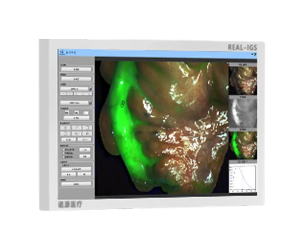
1. Ultra-low fluorescence detection limit: Real-time diagnosis of millimeter-sized tumors during surgery, decreasing detection limits to sub-millimeter levels.
2. Dual Quantitative Analysis Functions: ROI Value and Independent Spectral Digitization Quantitative Function provide objective numerical judgment.
3. Pseudo-color grading analysis system: Objectively presents the concentration gradient of the tracer in the target tissue.